The retina is the light-sensing tissue that lines the interior wall of the eye. Inside the main body of the eye is the vitreous gel, and the gel is adherent to the surface of the retina. As a normal aging process, the vitreous begins to degrade and consolidate, causing it to shrink, liquefy, and eventually separate from the retina. During this process, a mechanical pulling force is applied to the retina, causing stimulation that gives the sensation of flashing lights known as photopsia. When the gel is separated from the retina, it will cast a shadow that is seen as a floater and can take the shape of a speck, line, dot, circle, or spiderweb. The onset of flashes and floaters indicates an active process of posterior vitreous detachment from the retina.
In many patients, the process of posterior vitreous detachment will undergo its completion without symptoms; in those with symptoms, there is a 10-12% risk of developing a retinal tear. A tear in the retina can occur when the vitreous gel pulls on an area of a weak retina or in an abnormally adherent area. It is typically far in the peripheral retina if a tear occurs and will not affect the vision. However, fluid can migrate through a tear, causing the retina to detach from the wall of the eye.
Retinal detachments can cause vision loss and require surgery to treat. If the retinal tear is identified early, it can be treated to prevent a retinal detachment. The vitreous detachment can take several weeks to complete its course. During this process, there is an ongoing risk of developing a retinal tear, though the risk is highest at the onset and decreases with time.
While the process is a natural part of eye aging, specific conditions can accelerate the process. Patients with high myopia who have had trauma to the eye, undergone cataract extraction or other intraocular surgery, and have had YAG capsulotomy are at risk of having an acute posterior vitreous detachment. When the vitreous detaches secondary to one of these causes, there is an increased risk of developing a retinal tear.
A vitreous detachment causes not all floaters. The vitreous gel can consolidate and cast a shadow that causes floaters without detaching from the retina. These floaters can be present from a young age but are typically small and do not indicate any risk to the retina. Intraocular inflammation (Uveitis) can also cause floaters due to clumps of inflammatory cells inside the eye. Inflammation is typically associated with eye pain, sensitivity to light, and redness.
Not all flashes of light are caused by a vitreous detachment as well. Occasionally, photopsia can be caused by an ocular migraine. In this case, the retina is not being stimulated mechanically or by light, but a spasm of the blood vessels causes the sensation in the brain's visual cortex. These are typically bright, geometric, and multicolored but can also present as a dark spot. The standard pattern is for the lights to start small and increase in size over 20-30 minutes, then dissipate and can be associated with or without a headache.
It is difficult to differentiate the cause of flashing lights or floaters based on symptoms, and a thorough exam is indicated to evaluate any symptoms.
When the floaters first appear, they cast a shadow from being very close to the retina, causing the shadow to be very dark and dense; as time passes, the gel moves away from the retina, and the shadow becomes less dense. The floaters will never go away entirely, but as the darkness of the shadow decreases and time passes, they become more minor and less bothersome. The only reliable treatment is surgically removing the floaters with a Pars Plana Vitrectomy (PPV).
PPV is a surgical procedure that minimizes damage to the retina, by accessing the posterior segment of the eye and removing the vitreous. Small incisions are made in the wall of the eye. These incisions are positioned in the space behind the iris and front of the retina in the area of the Pars Plana.
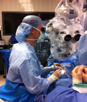
Surgery is performed in the operating room under sterile conditions
The surgery is performed in the operating room under sterile conditions. The procedure starts with a giant microscope to look through the pupil with special focusing lenses that provide a detailed and magnified view. Three incisions are made, and through one, a fluid infusion is placed, the second a light to provide illumination, and the third a vitreous cutter or another surgical instrument. Carefully, the vitreous is removed from the center of the eye, and the posterior vitreous is separated from the retina. The vitreous base is closely shaved to remove all vitreous gel possible. As the gel is being removed, the volume in the eye is replaced with a specially prepared, nutrient-rich, pH-balanced solution. Depending on the surgery, the eye may remain filled with this solution, and it will be slowly absorbed and replaced with the eye’s natural fluid. Other surgeries may require all fluid in the eye to be removed and a gas bubble placed in the eye to support the retina.
The most significant risk with any incisional surgery is the risk of infection; in the eye, this is known as endophthalmitis. Endophthalmitis is a severe condition that can lead to severe ocular damage and blindness. Great care is taken when performing eye surgery to minimize this risk, and this is why these procedures are performed under sterile conditions in the operating room. The eye is cleaned with betadine to kill surface bacteria, and antibiotics are used during and after surgery. New surgical techniques with small incisions have significantly decreased the risk of infection.
Additional risks with surgery include tissue damage that may result in retinal detachment, retina scarring, and elevated eye pressure (glaucoma). While all possible measures are taken to reduce these risks as much as possible, they can never be zero. It is crucial to weigh the risks with the benefits of the surgery when deciding on surgery.
As a consequence of removing the vitreous, cataract formation is accelerated. While all people will develop a cataract as a normal aging process, having vitrectomy surgery makes the cloudiness of the eye’s lens happen quicker. Cataracts are a treatable condition but require surgery to remove the lens and replace it with a clear lens implant.
Depending on the type of surgery, the eye may be filled with gas or silicone oil to hold the retina in place while healing. To keep these vitreous substitutes supporting the retina, the eye needs to be positioned facing down, allowing the gas or oil to float up to maintain contact with the retina. Different procedures require different lengths of time in the face-down position. Intraocular gas will resorb and be replaced by the eye’s natural fluid. Unique formulations are prepared to resorb at different rates, giving support for differing periods. Silicone oil does not resorb and will maintain its support until removed.
Pars plana vitrectomy is major surgery for the eye. During recovery, care must be taken to provide a suitable environment for the eye and retina to heal. It is essential to avoid any water in the eye for the first week after surgery to allow the incisions to heal. Limited physical activity is necessary for one month after surgery to give the incisions in the eye time to strengthen. The most crucial factor is the presence of gas or oil to support the retina. Time away from work should be taken as long as it is necessary to maintain the face-down position. Additionally, while a gas bubble is in the eye, vision will blur as light cannot pass through the gas, making work difficult and driving dangerous.
Request an appointment now