The tissue that senses the light inside the eye is the retina, and the retina's center is the macula. The macula is responsible for the central vision, and the cells are organized to give the central vision extra sensitivity. Inside the main body of the eye is the vitreous gel, and the gel is adherent to the retina's surface. As a normal aging process, the vitreous begins to degrade and consolidate, causing it to shrink and liquefy. This causes the vitreous gel to separate from the retina. During this process, force applied to the retina causes a mechanical pulling on the tissue, and in some people, this force can be too strong. The retina can be distorted, resulting in vitreomacular traction. The distortion of the retina can be very mild and not cause visual disturbance or be very severe with significant vision changes.
Most people with VMT have no symptoms, and the traction is seen on routine exams. While not specific for VMT, the most common symptom is a central blurry spot or fog. This may be experienced as haziness to the center of words while reading or in the center of the vision while the surrounding vision remains clear. Less commonly, but more specifically, there can be a bend or dip to straight lines or straight objects. This distortion is known as metamorphopsia. Since the condition affects the very center of the vision, the symptoms can be pretty disturbing.
Vitreomacular traction affects people of all types. Since the underlying process involves degeneration of the vitreous most people who experience VMT are in their 50s and 60s. Patients who have proliferative diabetic retinopathy or are very severely myopic can have a more severe form of the condition.
The basis of diagnosis is a thorough examination of the retina through a dilated pupil. The traction can be visualized, and any associated eye diseases were evaluated. Beneficial is the use of Optical Coherence Tomography (OCT) scanning. The OCT scans the retina and displays a cross-sectional picture that clearly shows the traction and can be used to measure the amount of adherence and level of distortion. The OCT scan can be compared to previous scans and follow the condition's progression.
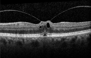
OCT of the macula reveals how the retina becomes distorted from vitreomacular traction
Typically the vitreous will detach from the retina naturally with time, and many patients can be observed to monitor the progress. In more severe cases, the vision may worsen, and treatment becomes necessary. For non-surgical treatment, there is ocriplasmin (Jetrea), an enzyme injected into the eye to break down the vitreous gel. In two extensive studies, the success of the injection was 26% in relieving the traction; alternatively, surgery can be performed. The Pars Plana Vitrectomy procedure is performed in the operating room under sterile conditions. Small incisions are made in the eye, and, using a microscope for viewing, tiny instruments are used to remove the vitreous gel that fills the inner eye, relieving the traction on the macula. If any epiretinal membrane tissue is present, it is removed from the retina's surface during the same procedure. A gas bubble may be placed in the eye, and head positioning is required. Not all patients will need this, and the decision is made during the surgery depending on findings during the procedure. The advantage of the surgery is that it is entirely successful at relieving the traction.
After the vitreous traction is relieved, the retina can regain its normal configuration, improving vision. Most people regain excellent vision, but some will have mild impairment. The recovery time is variable and can take several months to achieve the best vision possible.
Request an appointment now