Epiretinal membrane (ERM) can also be known as “macular pucker” or “cellophane retinopathy” and is scar tissue on the surface of the retina that causes wrinkling and distortion of the anatomy of the macula. Since the macula is responsible for central vision, a small change can cause significant visual problems. An ERM can be due to the normal aging process of the eye, retinal tears, prior surgery, eye trauma, or inflammation. When the retina is damaged, it will try to heal itself and release cells that form scar tissue like all tissues. Sometimes the scar-forming cells land on the retina's surface and begin to proliferate; Simultaneously, this tissue does not cause any direct damage to the retina. It can contract as it matures and distorts the structure of the retina.
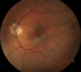
Epiretinal membranes can affect the macula, causing decreased central vision
As the retinal anatomy changes, the vision may begin to worsen. While not specific to changes from an ERM, the most common symptom is the vision becoming blurred or “smudged” and can occur in the central vision or near the center. More specifically, the vision remains clear to an ERM but becomes distorted. Straight lines such as door frames, window blinds, or highway stripes may develop waviness or bumps. A detailed examination needs to ensure an ERM is the cause of vision changes and not a more severe condition.
A thorough examination of the retina through a dilated pupil is necessary to diagnose an ERM. The epiretinal membrane can be seen on the retina's surface, and the amount of distortion can be assessed. A scan of the retina with an Optical Coherence Tomography (OCT) is often performed that allows the retinal layers to be analyzed and the ERM measured. It is also important to evaluate the eye for a potential membrane cause, as many conditions can cause permanent vision loss if not treated.
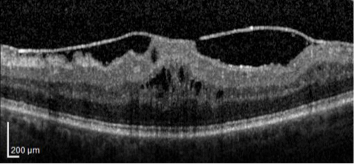
OCT scan of epiretinal membrane distorting the retinal structure
Initially, the membranes can be observed until the distortion, and visual compromise level becomes significant. The treatment for an epiretinal membrane is surgical removal with a Pars Plana Vitrectomy (PPV) and is performed in the operating room under sterile conditions. Small incisions are made in the eye, and, using a microscope for viewing, tiny instruments are used to remove the vitreous gel that fills the inner eye. After the gel is removed, high magnification is used to visualize the scar tissue, and specialized forceps carefully peel the scar tissue from the retina's surface. Depending on the nature of the membrane, the innermost layer of the retina, the internal limiting membrane, may also be removed simultaneously. Following surgery, the retinal distortion resolves, and the vision improves. The level of visual recovery is different for every patient and can take several months before maximum improvement is achieved.
Request an appointment now